Young Girl's Struggle with Back Pain Reveals Rare Spine Tumor
Key Takeaways
- A young girl experienced severe back pain and leg weakness due to a spine tumor.
- MRI scans revealed a large mass causing spinal cord compression.
- Urgent surgery was required to prevent permanent neurological damage.
Did You Know?
Introduction
A 10-year-old girl, previously healthy, experienced severe back pain for six months, which slowly worsened over time. Along with the pain, she started feeling numbness and weakness in her legs, causing her to struggle with walking and experience frequent falls. Her condition was severe enough to require a visit to the emergency department.
Initial Diagnosis
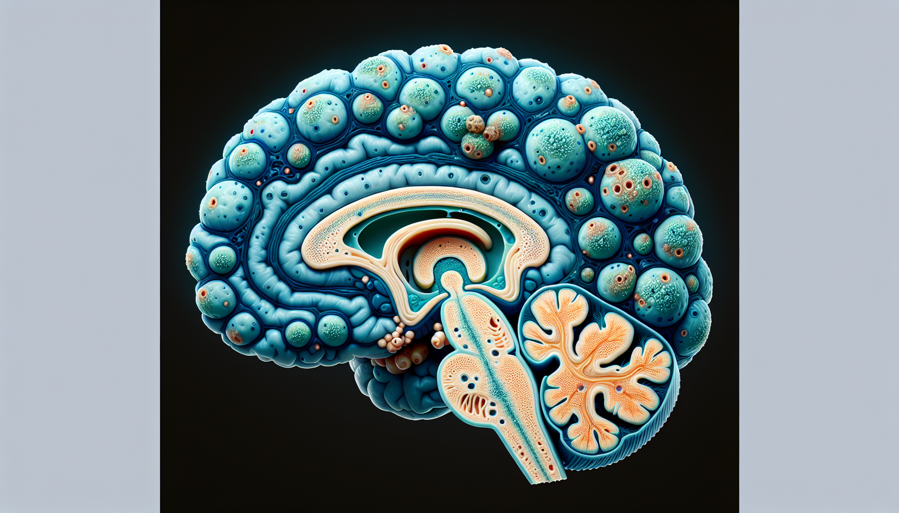
An MRI scan of her spine showed she had a large mass in her thoracic spine segment, specifically around the T10 vertebra, extending to nearby areas. This mass compressed her spinal cord, leading to her symptoms. The patient was admitted to the intensive care unit (ICU) for close monitoring and treatment with steroids aimed at reducing inflammation.
Challenges in Treatment
Treating this spine mass presented several challenges. First, the mass was growing rapidly, causing progressive myelopathy or spinal cord dysfunction. Without knowing exactly what type of tumor it was, doctors faced difficulties in planning the next steps. In cases with sudden declines in neurological function, quick decisions are necessary. Options vary depending on the nature of the mass, with some cases potentially requiring chemotherapy or radiation before surgery.
Emergency Surgery Decision
Given the rapid decline in her neurological function and the lack of immediate final pathology results, the medical team decided urgent surgery was necessary to prevent further damage. The risks of surgery include potential injury to the spinal cord, infection, and bleeding, but waiting could result in irreversible loss of function.
Surgical Procedure
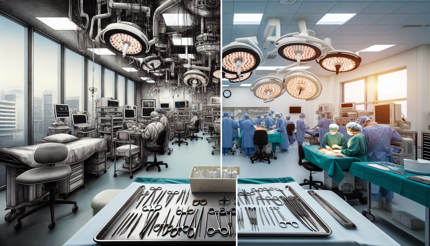
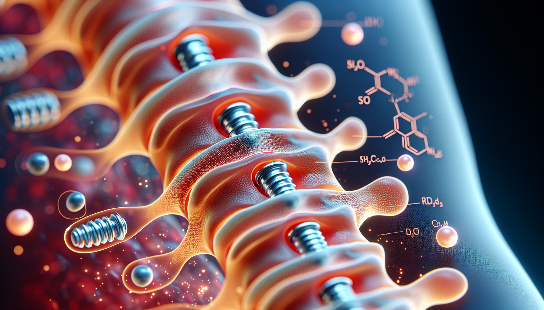
The girl underwent a major surgery that involved decompressing the spinal cord and stabilizing the spine with hardware. Surgeons removed part of the tumor and bone in a procedure that took many hours. They used specialized tools and techniques to minimize damage to surrounding structures.
Postoperative Care
After surgery, she was kept in bed and gradually started physical rehabilitation. Her strength and feeling in the legs began to improve, and follow-up care involved wearing a brace to support her spine while it healed.
Pathology Results and Diagnosis
The final pathology report identified the mass as an aneurysmal bone cyst (ABC), a benign but aggressive bone lesion that commonly affects young people. This diagnosis helped guide further treatment and follow-up.
Recovery and Follow-Up
By her six-week follow-up, she regained significant strength and was able to walk without assistive devices. She continued to show improvement at subsequent visits, and her brace was removed after three months. By six months, she had fully recovered and was allowed to return to some physical activities.
Aneurysmal Bone Cyst (ABC)
Aneurysmal bone cysts are rare, benign tumors that can grow aggressively and are more common in children and adolescents. They typically show up in the spine or long bones and can cause significant pain and neurological symptoms due to their size and location.
Treatment Options for ABC
There are several ways to treat ABCs, ranging from non-surgical methods like sclerotherapy and embolization to surgical removal. Complete surgical removal is often preferred to reduce the risk of recurrence, especially in cases where the cyst causes severe symptoms.


